

Anti-CANT1 Polyclonal Antibody
- Catalog number: PA11212
- Availability: In Stock
$168.00
- Ex Tax: $168.00
- Description: Anti-CANT1 Polyclonal Antibody (PA11212) is a rabbit polyclonal antibody detecting CANT1 in ELISA, IHC, WB. Suitable for Human, Mouse, and Rat.
Highlights:
• Affinity Purified — Minimal background and high purity for reliable results.
• Multi-Application — Validated across multiple applications.
• Multi-Species — Cross-reactive for translational research. - Host species: Rabbit
- Species reactivity: Human, Mouse, Rat
- Immunogen: E. coli - derived recombinant Human CANT1 (Ile107-Phe400).
- Form: Liquid
- Storage buffer: 0.01M PBS, pH 7.4, 50% Glycerol, 0.05% Proclin 300.
- Clonality: Polyclonal
- Isotype: IgG
- Applications: ELISA, IHC, WB
- Target: CANT1,Putative NF-kappa-B-activating protein 107,Apyrase homolog,Soluble calcium-activated nucleotidase 1,SCAN-1,SHAPY,Putative MAPK-activating protein PM09
- Purification: Purified by antigen affinity column.
- Stability and Storage: Use a manual defrost freezer and avoid repeated freeze thaw cycles. Store at 2 to 8°C for frequent use. Store at -20 to -80°C for twelve months from the date of receipt.
- Background: Soluble calcium-activated nucleotidase 1 (CANT1) is a ~44 kDa protein. Calcium-dependent nucleotidase with a preference for UDP. The order of activity with different substrates is UDP > GDP > UTP > GTP. Has very low activity towards ADP and even lower activity towards ATP. Does not hydrolyze AMP and GMP. Involved in proteoglycan synthesis.
References:
1. Smith, TM. et al. (2002) Archives of biochemistry and biophysics 406, 105-15. PMID: 12234496
2. Dai, J. et al. (2004) Cell 116, 649-59. PMID: 15006348
3. Yang, M. et al. (2004) Biochemistry 43, 9185-94. PMID: 15248776
4. Yang, M. et al. (2006) The Journal of biological chemistry 281, 28307-17. PMID: 16835225
5. Nizon, M. et al. (2012) Human mutation 33, 1261-6. PMID: 22539336 - Note: For research use only.
Images
-
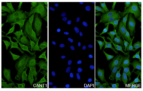 Immunofluorescence
Hela cells were fixed with 4% paraformaldehyde for 20 minutes, followed by blocking with 5% goat serum for 1 hour. The cells were then incubated with a primary antibody targeting CANT1 (HV161014) at a concentration of 26 ug/ml overnight at 4°C. Afterward, the cells were incubated with a secondary antibody, Goat Anti-Rabbit IgG (Alexa Fluor-488) for 50 minutes at room temperature. The localization of CANT1 was visualized (shown in green), and nuclear DNA was stained with DAPI (shown in blue).
Datasheet