
Anti-Human ATP6V1A Polyclonal Antibody
- Catalog number: PA11799
- Availability: In Stock
$168.00
- Ex Tax: $168.00
- Description: Anti-Human ATP6V1A Polyclonal Antibody (PA11799) is a rabbit polyclonal antibody detecting ATP6V1A in ELISA, IHC, WB. Suitable for Human, Mouse, Bovine, Chicken, and Pongo abelii.
Highlights:
• Affinity Purified — Minimal background and high purity for reliable results.
• Multi-Application — Validated across multiple applications.
• Multi-Species — Cross-reactive for translational research. - Host species: Rabbit
- Species reactivity: Human
- Immunogen: E. coli - derived recombinant Human ATP6V1A (Lys456-Asp617).
- Form: Liquid
- Storage buffer: 0.01M PBS, pH 7.4, 50% Glycerol, 0.05% Proclin 300.
- Clonality: Polyclonal
- Isotype: IgG
- Applications: ELISA, IHC, WB
- Target: ATP6V1A1, ATP6A1, VPP2, V-type proton ATPase catalytic subunit A, V-ATPase 69 kDa subunit, V-ATPase subunit A, ATP6V1A, Vacuolar proton pump subunit alpha, Vacuolar ATPase isoform VA68
- Purification: Purified by antigen affinity column.
- Stability and Storage: Use a manual defrost freezer and avoid repeated freeze thaw cycles. Store at 2 to 8°C for frequent use. Store at -20 to -80°C for twelve months from the date of receipt.
- Background: V-type proton ATPase catalytic subunit A (ATP6V1A) is a ~68 kDa protein. Catalytic subunit of the V1 complex of vacuolar(H+)-ATPase (V-ATPase), a multisubunit enzyme composed of a peripheral complex (V1) that hydrolyzes ATP and a membrane integral complex (V0) that translocates protons. V-ATPase is responsible for acidifying and maintaining the pH of intracellular compartments and in some cell types, is targeted to the plasma membrane, where it is responsible for acidifying the extracellular environment. In aerobic conditions, involved in intracellular iron homeostasis, thus triggering the activity of Fe(2+) prolyl hydroxylase (PHD) enzymes, and leading to HIF1A hydroxylation and subsequent proteasomal degradation. May play a role in neurite development and synaptic connectivity.
References:
1. van Hille, B. et al. (1993) The Journal of biological chemistry 268, 7075-80. PMID: 8463241
2. Vasanthakumar, T. et al. (2020) Trends in biochemical sciences 45, 295-307. PMID: 32001091
3. Miles, AL. et al. (2017) eLife 6. PMID: 28296633
4. Fassio, A. et al. (2018) Brain : a journal of neurology 141, 1703-1718. PMID: 29668857 - Note: For research use only.
Images
-
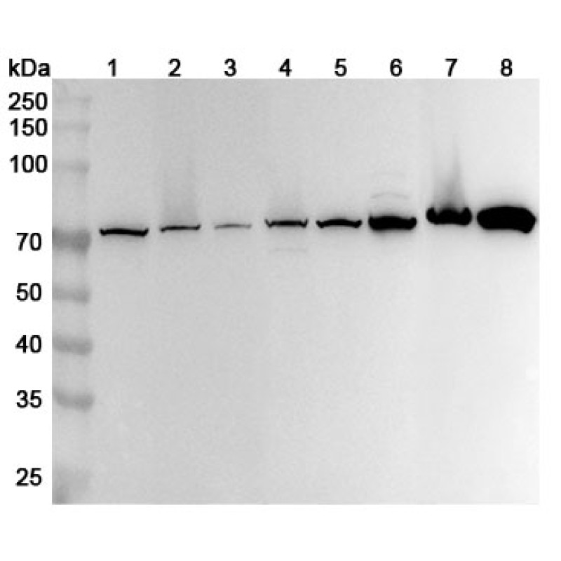
 Western blot
Western blot analysis was performed using anti-ATP6V1A polyclonal antibody at 1ug/mL on various samples.
Lane 1: HepG2 cell lysate
Lane 2: K562 cell lysate
Lane 3: U87 cell lysate
Lane 4: Daudi cell lysate
Lane 5: NIH3T3 cell lysate
Lane 6: Mouse brain lysate
Lane 7: Mouse kidney lysate
Lane 8: Rat brain lysate