

Anti-Human MSPR/Ron Reference Antibody (Narnatumab, RUO)
- Catalog number: BS10181
- Availability: In Stock
$176.00
- Ex Tax: $176.00
Description: Narnatumab (BS10181) is a research-grade recombinant antibody targeting CD136. Produced in mammalian cells with native-like glycosylation.
Highlights:
• Research Grade — For PK/PD studies, assay development, and ADA research.
• Native Glycosylation — Mammalian expression ensures native-like patterns.
Host species: Human
Isotype: IgG1-kappa
Species reactivity: Human
Immunogen:
Form: Liquid
Storage instructions:
Storage buffer: 0.01M PBS, pH7.4.
Concentration: 1mg/ml
Purity: >95% as determined by SDS-PAGE.
Clonality: Monoclonal
Applications: Research Grade Biosimilar
Target: CD136,Macrophage-stimulatingproteinreceptor,PTK8,MST1R,p185-Ron,RON,CDw136,MSPreceptor,Protein-tyrosinekinase8
Purification: Protein A/G purified from cell culture supernatant.
Endotoxin level: <1EU/mg, determined by LAL gel clotting assay
Expression system: Mammalian Cells
Stability and Storage: Use a manual defrost freezer and avoid repeated freeze-thaw cycles. Store at 4°C short term (1-2 weeks). Store at -20°C 12 months. Store at -80°C long term.
Alternative Names: IMCRON-8,RON8,CAS:1188275-92-4
Background: Macrophage-stimulating protein receptor (CD136/MST1R) is a ~152 kDa protein. Receptor tyrosine kinase that transduces signals from the extracellular matrix into the cytoplasm by binding to MST1 ligand. Regulates many physiological processes including cell survival, migration and differentiation. Ligand binding at the cell surface induces autophosphorylation of RON on its intracellular domain that provides docking sites for downstream signaling molecules. Following activation by ligand, interacts with the PI3-kinase subunit PIK3R1, PLCG1 or the adapter GAB1. Recruitment of these downstream effectors by RON leads to the activation of several signaling cascades including the RAS-ERK, PI3 kinase-AKT, or PLCgamma-PKC.
Note: For research use only. Not suitable for clinical or therapeutic use.
Highlights:
• Research Grade — For PK/PD studies, assay development, and ADA research.
• Native Glycosylation — Mammalian expression ensures native-like patterns.
Host species: Human
Isotype: IgG1-kappa
Species reactivity: Human
Immunogen:
Form: Liquid
Storage instructions:
Storage buffer: 0.01M PBS, pH7.4.
Concentration: 1mg/ml
Purity: >95% as determined by SDS-PAGE.
Clonality: Monoclonal
Applications: Research Grade Biosimilar
Target: CD136,Macrophage-stimulatingproteinreceptor,PTK8,MST1R,p185-Ron,RON,CDw136,MSPreceptor,Protein-tyrosinekinase8
Purification: Protein A/G purified from cell culture supernatant.
Endotoxin level: <1EU/mg, determined by LAL gel clotting assay
Expression system: Mammalian Cells
Stability and Storage: Use a manual defrost freezer and avoid repeated freeze-thaw cycles. Store at 4°C short term (1-2 weeks). Store at -20°C 12 months. Store at -80°C long term.
Alternative Names: IMCRON-8,RON8,CAS:1188275-92-4
Background: Macrophage-stimulating protein receptor (CD136/MST1R) is a ~152 kDa protein. Receptor tyrosine kinase that transduces signals from the extracellular matrix into the cytoplasm by binding to MST1 ligand. Regulates many physiological processes including cell survival, migration and differentiation. Ligand binding at the cell surface induces autophosphorylation of RON on its intracellular domain that provides docking sites for downstream signaling molecules. Following activation by ligand, interacts with the PI3-kinase subunit PIK3R1, PLCG1 or the adapter GAB1. Recruitment of these downstream effectors by RON leads to the activation of several signaling cascades including the RAS-ERK, PI3 kinase-AKT, or PLCgamma-PKC.
Note: For research use only. Not suitable for clinical or therapeutic use.
Images
-
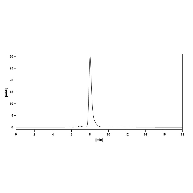
 SDS-PAGE
SDS-PAGE for Research Grade Narnatumab.
-
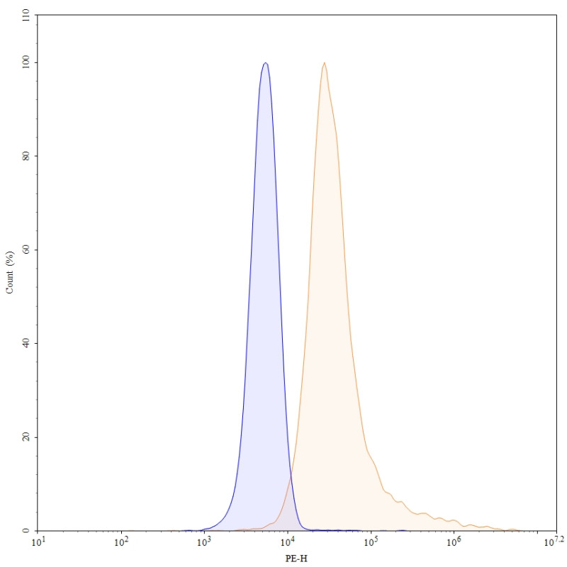
 Flow CytoMetry
Flow-cytometry using PE anti-human CD136 antibody. SW480 cells were stained with an irrelevant antibody (Blue Histogram) or an PE anti-human CD136 monoclonal antibody (Catalog: HC609016, Yellow Histogram) at a concentration of 5 µg/ml for 30 mins at RT. After washing, and cells analysed on a NovoCyte Flow Cytometer.